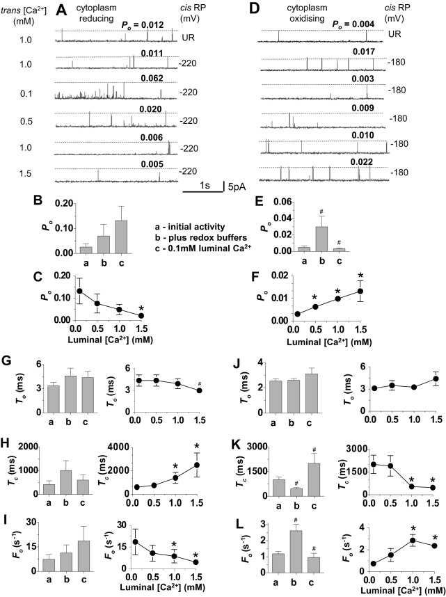
Fig. 3.
Effect of cytoplasmic and luminal GSH∶GSSG redox buffers on the response of RyR2 channels to changes in luminal [Ca2+]. In this and subsequent figures, the redox potential (RP) in all luminal solutions was −180 mV, the cytoplasmic [Ca2+] was 1.0 µM, channel activity was recorded at +40 mV, channel opening is upwards and open probability (Po) values for each recording are shown above the broken line. (A–C,G–I) Channels exposed to cytoplasmic solutions having a reducing redox potential of −220 mV (n = 10). (D–F,J–L), channels exposed to cytoplasmic solutions having a more oxidised redox potential of −180 mV (n = 15). (A,D) Descending from the upper trace, the data show initial unbuffered redox (UR) activity with 1.0 mM luminal Ca2+, activity after addition of GSH∶GSSG with 1.0 µM luminal Ca2+, activity after perfusion with 0.1 mM Ca2+ luminal solution and replacement of GSH∶GSSG, then activity when luminal [Ca2+] was increased stepwise to 0.5 mM, 1 mM and 1.5 mM. Po values for each recording are shown. (B,E) Mean Po determined (a) for initial activity with 1 mM luminal Ca2+ and then (b) after adding GSH∶GSSG buffers and (c) after lowering luminal [Ca2+] to 0.1 mM. (C,F) Mean Po after stepwise increases in luminal [Ca2+] from 0.1 mM to 1.5 mM. (G–L) Mean gating parameter values. (G,J) Mean open time (To); (H,K) mean closed time, (Tc); (I,L) mean frequency of opening (Fo). The bar graphs show mean parameter values (a) for initial activity with 1 mM luminal Ca2+, (b) after adding GSH∶GSSG buffers and (c) after lowering luminal Ca2+ to 0.1 mM. The line graphs are plots of mean parameter values as a function of luminal [Ca2+]. Data are shown as the mean±s.e.m.; #P<0.05 (versus the preceding condition); *P<0.05 (versus the mean value with 0.1 mM Ca2+).

