
Figure 2. Oligomerization of the Dam1 complex enhances microtubule attachment strength.
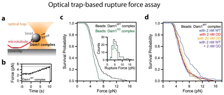
(a) Diagram of the optical trap assay. (b) Example trace of applied force versus time from the rupture force assay. Beads are subjected to a ~2.5-pN test force prior to initiation of the force ramp (~0.25 pN·s−1), which begins at t = 0 s. Dashed line indicates the rupture force from this event. (c) Survival versus force curves for beads coated with wild-type (WT, black trace, n = 120) or oligomerization-deficient (OD, green trace, n = 69) Dam1 complexes coupled to microtubule tips. Inset: same data represented in survival curves but re-plotted as rupture force histograms. (d) Survival versus force curves for beads coated with Dam1WT complex alone (black trace, reproduced from c) or in the presence of additional Dam1 complex free in solution: with 2 nM Dam1WT complex, blue, n = 106; with 2 nM Dam1OD complex, red, n = 86; with 20 nM Dam1OD complex, orange, n = 89; with 2 nM Dam1WT and 2 nM Dam1OD complex, purple, n =109.