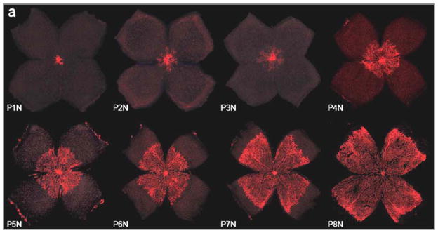
Figure 1.

Retinal vessels (red) grow in a centrifugal pattern from the optic nerve head towards the periphery of the reina, the ora serrata. In the mouse retina, retinal vascular growth occurs postnatally between postnatal day (P) 1 and 8 under normoxic (N) conditions (referred to as P1N – P8N in the Figure). In normal human development, Retinal vascularization occurs in utero and the retina is fully vascularized at term. However, when development is disrupted by preterm birth retinal vascular development can be severely suppressed or developed vessels can regress leading to ROP. Image reprinted with permission from [3] (Copyright holder: Association for Research in Vision and Ophthalmology).
