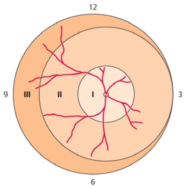
Figure 2.

Retinal zones used for grading of ROP. Zone I describes a circle around the optic nerve head (ONH) with a radius of twice the distance from ONH to fovea. Zone II is a circle around the ONH reaching the nasal periphery (at the 3 o’clock position in the image). Zone III is the remaining temporal crescent-shaped retina. Image reproduced with permission from [6].
