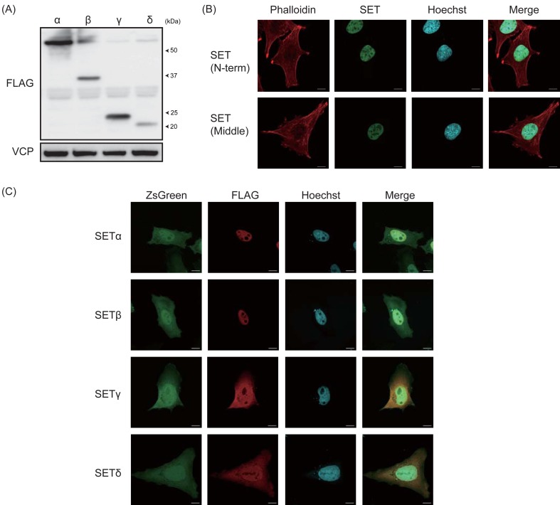
Fig. 4.
Expression and localization of canine SET isoforms. (A) HEK293T cells were transfected to express FLAGx3-tagged canine SET isoforms, and protein expression was analyzed by immunoblotting. VCP was used as a loading control. (B) The localization of endogenous SET in CMeC1 cells was examined by immunofluorescent staining using anti-SET (N-term) and anti-SET (Middle) (Green). Phalloidin (Red) and Hoechst (Blue) were used to stain F-actin and the nucleus. (C) HeLa cells were transfected to express FLAGx3-tagged canine SET isoforms and ZsGreen, and the localization of SET isoforms was determined by immunofluorescent staining using an anti-FLAG antibody (Red). Hoechst (Blue) was used to stain the nucleus.

