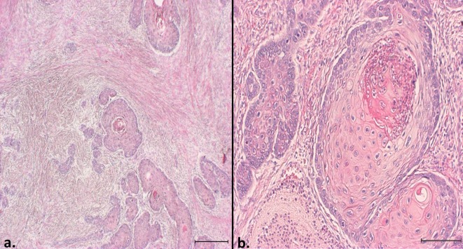
Fig. 3.
Histopathological features of a squamous cell carcinoma in a capybara. HE. a) The lesion comprised islands of invasive squamous epithelial tumor cells with a severe desmoplastic reaction. Bar=500 µm. b) The tumor cells showed various degrees of keratinization with keratin pearls. Inflammatory cells frequently infiltrated many of the keratin pearls. Bar=200 µm.

