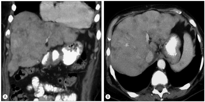
Figure 3.
Pseudocirrhosis. Fifty six-year-old female underwent multiple cycles of chemotherapy for metastatic liver disease from breast cancer. Coronal (A) and axial (B) contrast enhanced CT images of the abdomen show a macronodular liver with fibrosis following completion of therapy. Patient had features of early hepatic decompensation.

