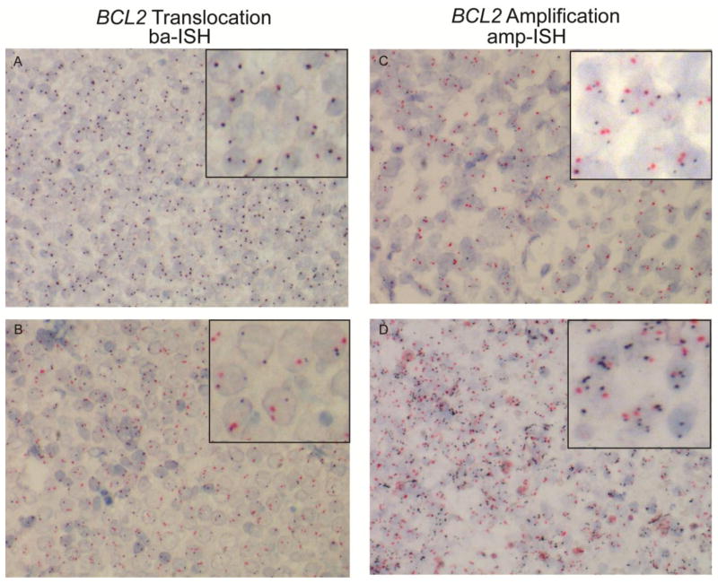
Fig. 2. BCL2 Dual ISH assays for detection of gene translocation and amplification.
Representative DLBCL FFPET sections stained with a break-apart probe set (A and B) specific for the 3′ and 5′ end of the BCL2 gene or an amplification probe set specific for chromosome 18 centromere and the BCL2 gene (C and D) shown as red and black signals, respectively. Overlapping red and black signals indicates the absence of translocation (A), while a “free”, non-overlapped red signal indicates translocation (B). For the amplification ISH, more than 2 black signals in the presence of 2 red signals indicates amplification. Inset is a portion of the section at high power. 400X magnification.

