
FIG. 2.
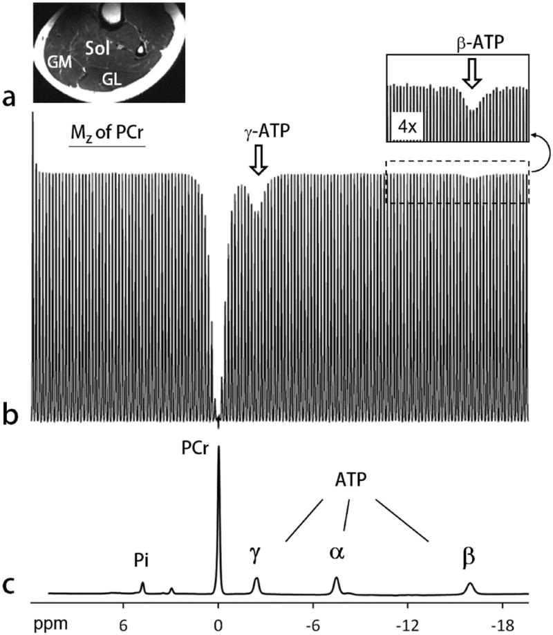
EKIT spectrum of human calf skeletal muscle. a: A typical T2w MR image acquired from human calf using a partial volume coil. Abbreviations: Sol, soleus; GM, gastrocnemius medial; GL, gastrocnemius lateral. b: The EKIT spectrum illustrated here reflects the intensity of PCr signal plotted as a function of frequency of the inversion pulse (150 different frequencies) acquired from calf of a 25 yr healthy female with a BMI = 22.2 kg/m2. Other 31P MRS acquisition parameters: TR 5 second, td 1 second, scan time 12.5 min. Note that PCr magnetization is attenuated after inversion of either γ ATP (white arrow) or β ATP (insert). c: A conventional 31P MR spectrum acquired showing the full chemical shift region of calf muscle 31P resonances, with the chemical shift aligned with that of sweeping inversion pulse used in the EKIT spectrum b.