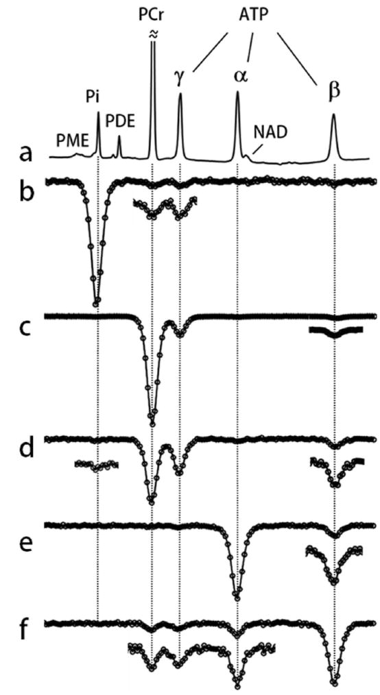
FIG. 4.

EKIT spectra of resting human calf muscle. a: A conventional 31P MR spectrum and (b-f) EKIT spectra with observation resonance at b: Pi, c: PCr, d: γ-ATP, e: α-ATP, and f: β-ATP acquired from resting human calf muscle at td = 1 s. These spectra represent averages from 7 subjects. In some spectra, small exchange peaks were enlarged 3-fold.
