
Figure 2.
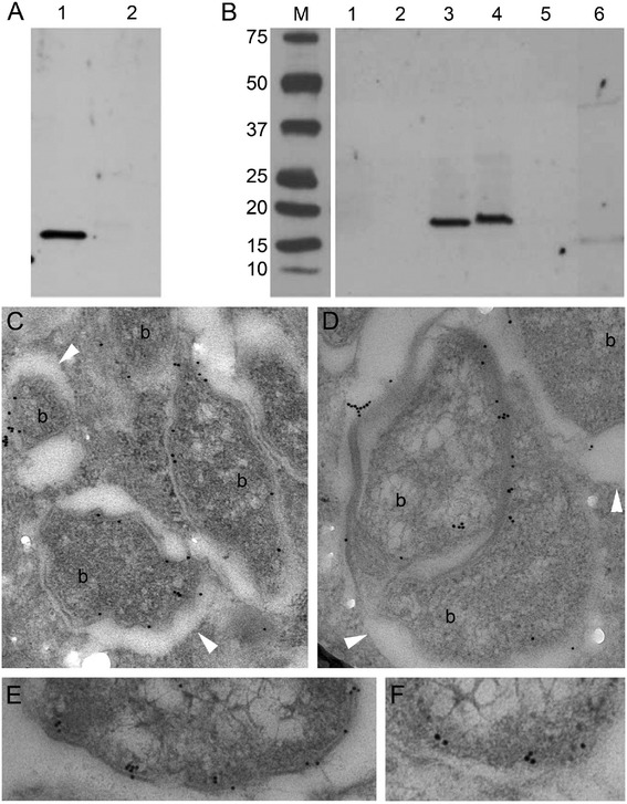
Localisation of PAL on Wolbachia membranes. A, B – Western immunoblot detection of wPAL protein in B. malayi (A) and C6/36 mosquito cell line fractional (B) samples. A. Protein samples of B. malayi infected wBm (1) and B. malayi after six weeks tetracycline treatment (2). B. Protein samples of infected and non-infected C6/36 cells cultivated in vitro: 1 – total protein extract of non-infected cells, 2 – membrane fraction of non-infected cells, 3 – total protein extract of cells infected with wAlbB, 4 – membrane fraction of infected cells, 5, 6 – cytoplasmic fractions of non-infected and infected cells, respectively. M – marker, kDa. C-F – Immuno-transmission electron microphotographs showed localisation of PAL on the bacterial membrane in cytoplasm of adult B. malayi infected with wBm. b – bacteria. Arrow head indicates membrane of vacuole containing Wolbachia.