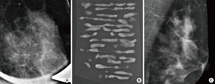
Figure 1.
A 45-year-old woman with ductal carcinoma in situ. (A) Magnification view of mediolateral mammography reveals clustered pleomorphic calcifications measuring 11 mm at the longest dimension in left upper central breast. Vacuum-assisted breast biopsy was performed with 11-gauge needle and the localizing clip was placed. (B) Radiography of the vacuum-assisted breast biopsy specimens revealed calcification and the diagnosis was atypical ductal hyperplasia. (C) Mediolateral mammography of the left breast obtained after 1 week shows localizing clip without evidence of residual calcifications. After surgery, the pathologic diagnosis was ductal carcinoma in situ.

