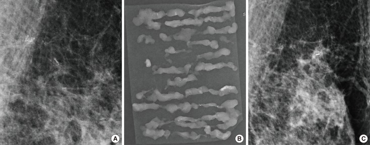
Figure 2.
A 55-year-old woman with ductal carcinoma in situ. (A) Magnification view of mediolateral mammography reveals linear distributed linear branching calcifications measuring 18 mm at the longest dimension in left upper medial breast. Vacuum-assisted breast biopsy was performed with 11-gauge needle and the localizing clip was placed. (B) Radiography of the vacuum-assisted breast biopsy specimens revealed calcification and the diagnosis was atypical ductal hyperplasia. (C) Mediolateral mammography of the left breast obtained after 1 week shows localizing clip with remaining calcifications. After surgery, the pathologic diagnosis was ductal carcinoma in situ.

