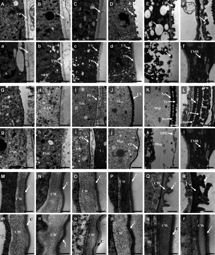
Fig. 4.
Comparison of anther and pollen between the wild type and osabcg15 by transmission electron microscopy. Ba bacula, C cuticle, CW cell wall, DMsp degenerated microspore, DPE degenerated prim-exine, EX exine, Msp microspore, Ne nexine, PMC pollen mother cell, PE prim-exine, Te tectum, Tds tetrads, TMR tapetum and microspore residue, TW tapetum cell wall, Ub Ubisch, V vacuole. Bars 1 μm in A–L, and a–l; 200 nm in M–R, and m–r. Cross-sections of the wild-type tapetum at stages 7 (A), 8a (B), 8b (C), 9 (D), 10 (E), and 11 (F). Cross-sections of the osabcg15 tapetum at stages 7 (a), 8a (b), 8b (c), 9 (d), 10 (e) and 11 (f). G–L The pollen exine development of the wild type from stages 7–11. g–l Defective pollen exine development of osabcg15 from stages 7–11. M–R Outer region of anther epidermis in the wild type from stages 7–11. m–r Outer region of the anther epidermis in the osabcg15 mutant from stages 7–11

