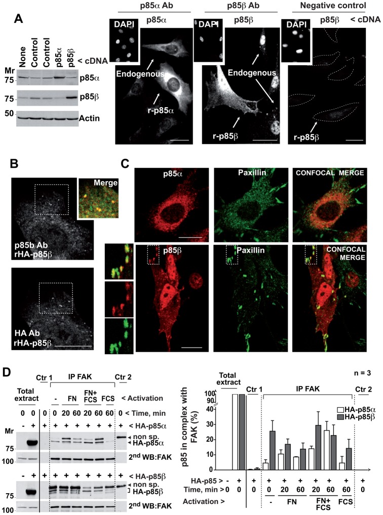
Fig. 1. p85β localizes at adhesion plaques and associates with FAK.
(A) NIH3T3 cells, untransfected or transfected with cDNA encoding p85α or p85β (48 h), were tested in Western blot (WB). Other cells were examined by immunofluorescence (IF) using p85α- or p85β (K1123)-specific antibodies. Insets show DAPI staining for nuclear DNA. To control K1123 specificity, some of the cells that were stained with the antibody (Ab), were preincubated with antigenic peptide. A dashed line outlines the cell membrane; transfected cells are indicated. (B) NIH3T3 cells transfected with HA-p85β cDNA were stained simultaneously with K1123 polyclonal Ab and an anti-hemagglutinin (HA) mAb; the inset shows a magnification of the indicated region. (C) NIH3T3 cells were transfected as above and tested in IF by simultaneous staining of paxillin and p85α or p85β. Insets show magnifications. (D) NIH3T3 cells were transfected with cDNA encoding HA-p85α or HA-p85β (48 h), then incubated without serum (16 h) and activated with 10% serum or in FN-coated wells with and without serum for 20 or 60 min. Cell extracts (500 µg) were immunoprecipitated with anti-FAK Ab. Negative controls included extract + protein A (Ctr 1) and protein A + Ab (Ctr 2); Extracts (50 µg) and immunoprecipitates were analyzed in WB using anti-HA Ab. Loading was controlled in WB using anti-FAK Ab. The graph shows HA signal intensity relative to HA-p85 expression in whole cell extract, considered 100% (mean ± s.d.; n = 3). Bar = 12 µm.

