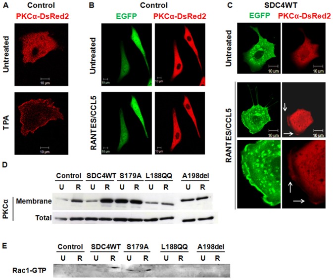
Fig. 5. RANTES/CCL5 induced co-localization of SDC-4 and PKCα at the cell membrane and Rac1 activation.
(A–C) HUV-EC-C were co-transfected with PKCα-DsRed2 plasmid and with either GFP plasmid (control, panels A and B) or GFP-SDC4WT (SDC4WT, panel C). They were incubated or not with (A) 0.5 µM TPA or with (B,C) 3 nM RANTES/CCL5 for 15 minutes and analyzed under live confocal microscopy. Membrane localization of SDC-4 (green) and PKCα (red) was indicated with white arrows. (×400). (D) HUV-EC-C transfected with GFP plasmid (control) or with SDC4WT-GFP (SDC4WT) or with mutated SDC-4 constructs (S179A, L188QQ, A198del) were stimulated or not (U) by 3 nM RANTES/CCL5 (R). After cell fractionation, the amount of PKCα in membrane of total fraction was evaluated by western blot. (E) HUV-EC-C transfected with GFP plasmid (control) or with SDC4WT-GFP (SDC4WT) or with mutated SDC-4 constructs (S179A, L188QQ, A198del) were stimulated or not (U) by 3 nM RANTES/CCL5 (R). Rac1-GTP activity was determined by pull down assay and analyzed using specific Rac1-GTP antibodies by western blot. Scale bars: 10 µm.

