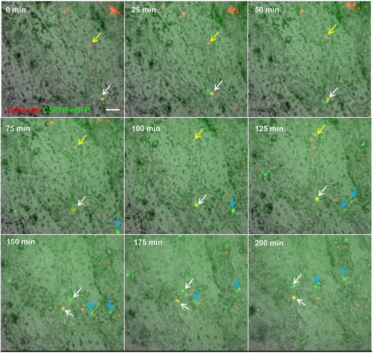
Fig. 4.

Macrophages associated with the embryonic vasculature are highly motile and phagocytic, and undergo local division. Time-lapse imaging of region above the vitelline artery near the embryo proper. The aorta of CSF1R-eGFP embryos was injected with Texas Red-labelled zymosan 1 h prior to the beginning of imaging. Most zymosan particles adhered to the blood vessel walls (yellow arrows). eGFP+ macrophages are highly motile. Between 100 and 125 min from the start of filming, a zymosan particle (yellow arrow) becomes associated with a macrophage; this macrophage re-enters the circulation, removing the zymosan particle by 150 min. At 0 min, a zymosan particle is contained within a macrophage (white arrow); from 0-75 min this macrophage is both motile and exhibits changes in morphology. At 100 min, this macrophage (white arrow) no longer exhibits movement and does not extend any cellular processes. A similar macrophage without a phagocytised zymosan particle (blue arrow) exhibits identical behaviour. At 100-150 min, both undergo division (white and blue arrows), and daughter cells resume active patrolling of the vasculature. Scale bar: 50 µm.
