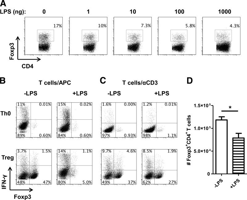
Figure 8. LPS inhibits Foxp3 Tregs.
(A) CD62L+CD4+ CBir1 CD4 T cells were cocultured with irradiated splenocytes with CBir1 peptide and 5 ng/ml TGF-β to induce Foxp3 induction. Cells were also treated with or without varying concentrations of LPS and analyzed by flow cytometry after 5 days. (B) CD62L+CD4+ CBir1 CD4 T cells were cocultured with irradiated splenocytes (APC) with CBir1 peptide in the absence of polarization (Th0) or with 5 ng/ml TGF-β (Treg). Cells were also treated with or without LPS (1000 ng) and analyzed by flow cytometry after 5 days. (C) CD62L+CD4+ T cells were cultured with plate-bound αCD3 (1 μg) and αCD28 (2 μg/ml) in the absence of polarization (Th0) or with 5 ng/ml TGF-β (Treg). Cells were also treated with or without LPS (1000 ng) and analyzed by flow cytometry after 5 days. (D) Bar graph reflects total numbers of Foxp3+ cells generated in culture of C and represents mean ± sem. *P < 0.05, n = 4. FACS plots are gated upon live CD4+ cells.

