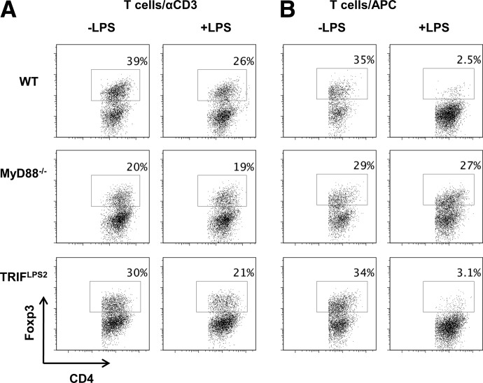
Figure 9. LPS partially inhibits Treg generation through MyD88 signaling on CD4 cells and APCs.
(A) MyD88−/−, TRIF−/−, or WT CD4 cells were cultured with plate-bound αCD3 (1 μg) and αCD28 (2 μg/ml) with 5 ng/ml TGF-β (Treg) in the presence or absence of LPS (1 μg) and analyzed by flow cytometry after 5 days. (B) MyD88−/−, TRIF−/−, or WT irradiated splenocytes (APC) were cultured with WT CD4 cells with plate-bound αCD3 (1 μg) and 5 ng/ml TGF-β in the presence or absence of LPS (1000 ng) and analyzed by flow cytometry after 5 days. FACS plots are gated upon live CD4+ cells.

