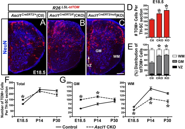
Fig. 4.
The number and distribution of Ascl1 lineage glial cells into the GM and WM are altered in Ascl1 CKO and Ascl1 KO spinal cords. Immunofluorescence on thoracic sections of E18.5 Ascl1CreERT2/+;R26LSL-tdTOM control, Ascl1CreERT2/FL;R26LSL-tdTOM (Ascl1 CKO) or Ascl1CreERT2/null;R26LSL-tdTOM (Ascl1 KO) mice treated with tamoxifen at E14.5. (A-E) NeuN delineates the boundaries between VZ, GM and WM. The number and distribution of tdTOM+ cells are increased in the GM of Ascl1 CKO and Ascl1 KO at E18.5. (F,G) Number of tdTOM+ cells is altered in the GM and WM of Ascl1 CKO into postnatal stages. Number of spinal cords analyzed: E18.5, N=4 control, N=3 Ascl1 CKO and Ascl1 KO; P14, N=3 control and Ascl1 CKO; P30, N=2 control and Ascl1 CKO. *P<0.005 indicates statistical significance using linear regression analysis. Values in graphs are mean±s.e.m. Scale bar: 100 μm.

