Fig. 8.
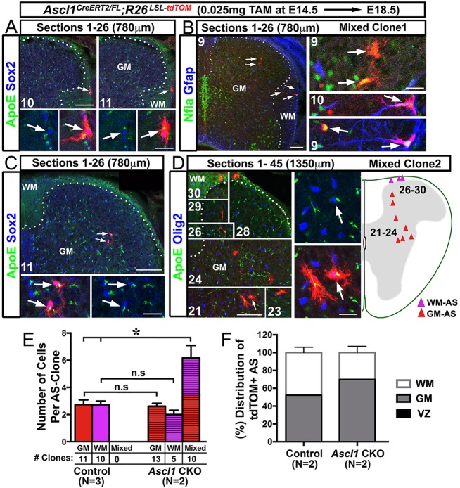
The proliferation and distribution of astrocyte clones in the GM and WM are altered in Ascl1 CKO spinal cords. Immunofluorescence on spinal cord sections of Ascl1CreERT2/FL;R26LSL-tdTOM mice treated with a low dose of tamoxifen (0.025 mg/40 g body weight) at E14.5. (A,C) Consecutive sections showing the presence of WM-only (A) or GM-only (C) labeled astrocyte clones found at the center of two separate 780 μm spinal cord portions. (B,D) Consecutive sections showing the presence of two mixed (GM and WM) astrocyte-labeled clones that are found separately in a 780 μm or 1350 μm spinal cord segment. Note that the number of cells in the mixed clones is increased compared with GM-only or WM-only clones. Dotted lines delineate the GM/WM boundary. Arrows indicate labeled AS noted in both low- and high-magnification images. (E) Number (mean±s.e.m.) of cells per AS clone. The number of each type of clone is also shown. (F) Distribution of sparsely labeled tdTOM+ AS cells in the VZ, GM and WM between control and Ascl1 CKO littermates. *P<0.005, Student's t-test; n.s, not significant. Scale bars: 100 μm for low-magnification images; 25 μm for high-magnification insets.

