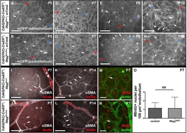
Fig. 7.
Following endothelial Rbpj deletion from birth, brain AV connections are abnormal by P8, lack αSMA and do not proliferate. (A-H) Whole-mount timecourse images showing brain AV connections (arrows). At P8, RbpjiΔEC AV connections (F) appear different than in controls (E). For each time point, N=3 controls, N=3 mutants, with at least seven fields imaged from each brain. (I-L) Whole-mount αSMA immunostaining. Arrows point to AV connections, which lacked αSMA staining in both control and mutant. For P7, N=4 controls, N=3 mutants; for P14, N=3 controls, N=3 mutants. (M-O) BrdU incorporation in ECs from P5-P7 was unchanged in mutant (N) versus control (M) brain (as quantified in O). Arrow indicates EC nucleus. P=0.9752, according to Student's t-test. N=4 controls (29 fields), N=5 mutants (33 fields). a, artery; v, vein. Scale bars: 100 μm in A-L; 25 μm in M,N.

