Fig. 2.
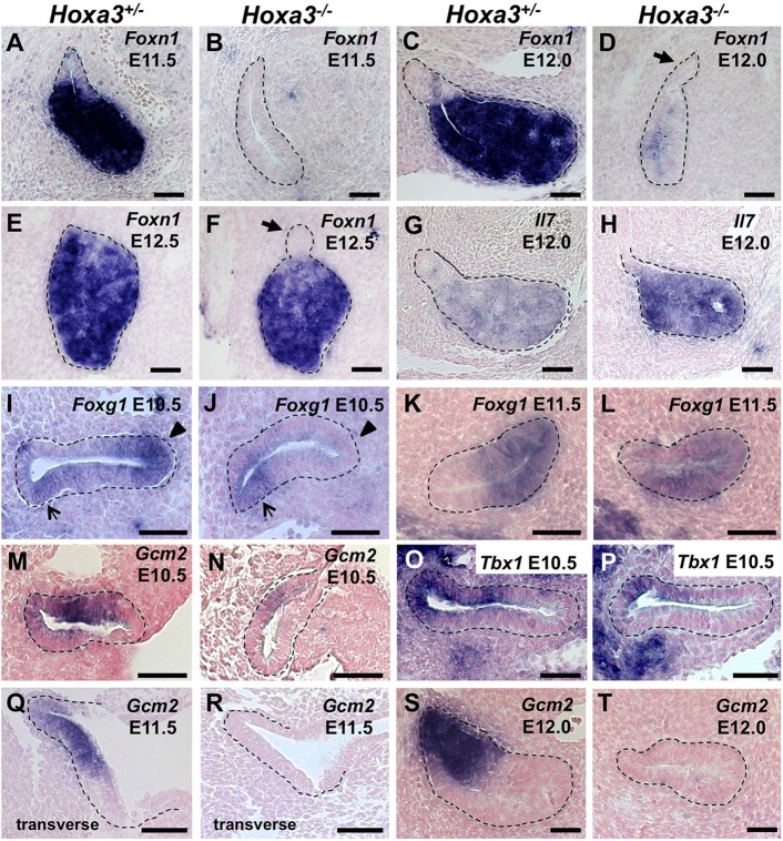
Foxn1, Il7, Foxg1, Gcm2, and Tbx1 are mis-expressed in Hoxa3−/− mutants. ISH of paraffin sections from Hoxa3+/− and Hoxa3−/− embryos. (A-F) Sagittal sections at E11.5 (n=3), E12.0 (n=2) and E12.5 (n=1) were stained with a Foxn1 riboprobe. Arrows indicate a group of Foxn1-negative cells attached to the thymus at E12.0 and E12.5. (G,H) Sagittal sections at E12.0 were stained with an Il7 riboprobe (n=2). (I-L) Sagittal sections at E10.5 (n=2) and E11.5 (n=2) were stained with a Foxg1 riboprobe. Foxg1 expression in the Hoxa3−/− is absent in the thymus domain (arrowheads) but present in the dorsal-posterior domain at E10.5 (arrows). Sagittal sections at E10.5 (n=4) (M,N), transverse sections at E11.5 (n=3) (Q,R) and sagittal sections at E12.0 (n=2) (S,T) were stained with a Gcm2 riboprobe. (O,P) Sagittal sections at E10.5 (n=2) were stained with a Tbx1 riboprobe. In all sections, anterior is towards the top of the image, and the same side is presented per stage and gene. The third pharyngeal pouch is outlined in all panels. Scale bars: 40 µm.

