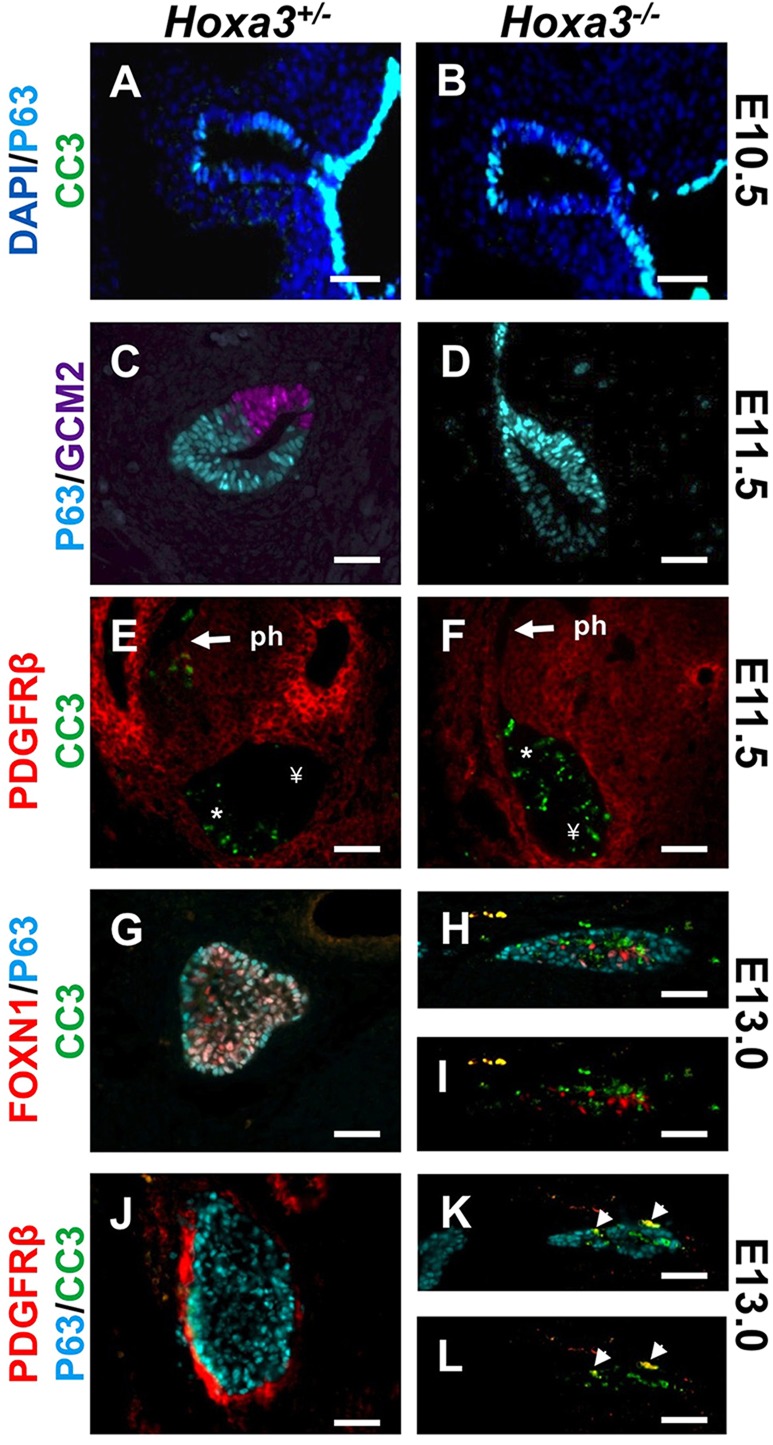
Fig. 3.

The parathyroid and thymus are lost in Hoxa3−/− mutants. Immunostaining of sagittal E10.5 (n=2) (A,B), E11.5 (n=3 each set) (C-F) and transverse E13.0 (n=3 each set) (G-L) sections of the Hoxa3+/− and Hoxa3−/− thymus with antibodies against cleaved caspase-3 (CC3), p63, GCM2, PDGFRβ and FOXN1. DAPI stains nuclei. *, the region within the primordium that normally undergoes apoptosis during separation from the pharynx. ¥, the posterior-dorsal region that does not normally undergo apoptosis. Arrows indicate the thymus-pharynx (ph) attachment domain that normally undergoes apoptosis. Arrowheads indicate co-labeling of PDGFR and CC3. Scale bars: 40 µm.
