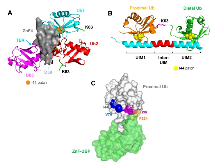
Figure 4.
Recognition of specific Ub chains by UBD-containing proteins.
(A) A20 ZnF4 domain (grey) recognizes K63-linked triubiquitin (PDB: 3OJ3). This occurs through the interaction of the I44 hydrophobic patch (orange sphere) of Ub1 (cyan), aspartate-58 on the 50s loop (yellow) of Ub2 (red) and the TEK-box of Ub3 (magenta). The positions of the putative K63 linkages are indicated [53]. (B) Crystal structure of the tandem UIMs (cyan) on Rap80 protein in complex with the extended K63-linked di-Ub chain (PDB: 3A1Q). Two UIM domains are separated by a 7 residue helix (red) for optimal recognition of the I44 patch (yellow spheres) on the proximal (orange) and distal (green) Ubs [58]. (C) ZnF-UBP (green) from the deubiquitinating enzyme IsoT recognizes and binds to the C-terminal glycine of a proximal Ub (grey) (PDB: 2G45). This occurs through the interaction of L8 and I36 of the Ub (magenta) and F224 of the ZnF-UBP (yellow) [64]. Copyright (2006), with permission from Elsevier.

