
Fig 2.
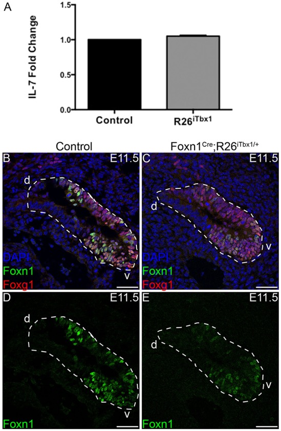
Ectopic TBX1 does not affect expression of IL7 or FOXG1 in the ventral 3rd pp. (A) Real-time quantitative PCR analysis shows equivalent Il7 mRNA levels in the 3rd pp dissected from E11.5 control or Foxn1Cre;R26iTbx1/+ embryos (n=6 control and n=6 Foxn1Cre;R26iTbx1/+). (B) Representative immunohistochemical stain showing a sagittal section of E11.5 3rd pp. FOXG1 (red) and FOXN1 (green) are co-expressed in the ventral (v) domain; DAPI (blue). (C) Representative immunohistochemical stain of a sagittal section of the mutant 3rd pp showing abundant FOXG1 in the ventral domain despite the severe reduction in FOXN1. (D,E) Single-color images of FOXN1 staining. n=4 control and n=4 Foxn1Cre;R26iTbx1/+ 3rd pps). Scale bars: 50 μm. d, dorsal.