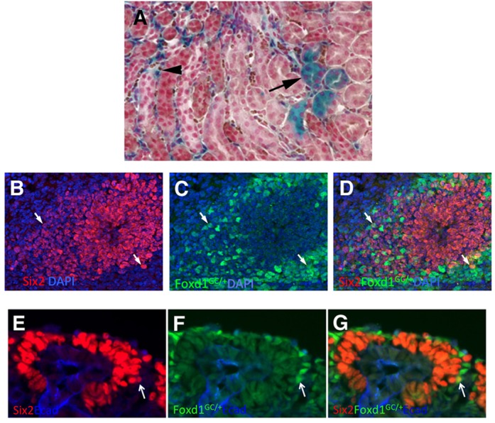
Fig. 2.

Cells expressing both Foxd1 and Six2. Cells expressing both Six2 and Foxd1 protein were present, but rarer than predicted by the transcriptional profiling. (A) A Foxd1-Cre transgenic mouse was mated to a floxed stop Rosa26-lacZ reporter mouse, with the resulting kidneys primarily showing stromal cell labeling, as expected (arrowhead), but with some tubular nephron epithelial cells also labeled (arrow). The counterstain used was nuclear Fast Red. (B-D) E11.5 kidney immunostained for Six2 (red) and Foxd1. The expression of Foxd1 was detected using a Foxd1-GFP-Cre knock-in mouse and GFP antibody. Two positive cells near the periphery of the Six2-positive population are marked with arrows. (E-G) E15.5 Foxd1-GFP-Cre knock-in embryos were immunostained for Six2 and GFP. A rare double-labeled cell, again at the periphery of the Six2-positive cells, is marked with an arrow.
