Fig. 1.

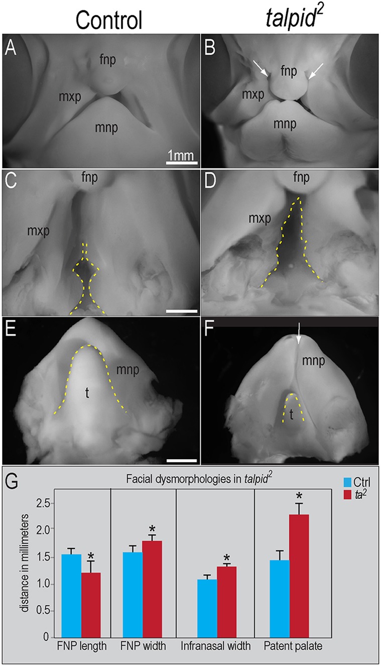
Growth and development of the facial prominences is disrupted in talpid2 mutants. Day 7 control (n=10) and talpid2 (n=4) embryos. (A,B) Frontal views of facial prominences; frontonasal (fnp), maxillary (mxp) and mandibular prominences (mnp). Bilateral clefting in talpid2 embryos (white arrows in B). (C,D) Palatal views show increased patency of both the primary and secondary palate of talpid2 embryos (dotted yellow line). (E,F) Dorsal view of the mnp. The mnp of talpid2 embryos is clefted (white arrow in F) and exhibits hypoglossia (dotted yellow line). (G) Quantitative analysis of fnp and mxp growth. For fnp length, *P<0.02; for fnp width, *P<0.02; for infranasal width, *P<0.04; for patent palate, *P<0.02. t, tongue. Data are mean±s.e.m. Scale bars: 1 mm (A-F).
