Fig. 7.

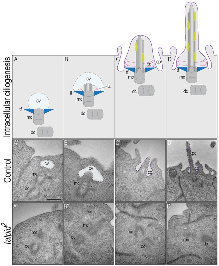
Intracellular ciliogenesis is disrupted in talpid2 embryos. Hypothesized steps of intracellullar ciliogenesis. (A) A ciliary vesicle (cv) binds to the distal end of the mother centriole (mc), via associations with transition fibers (tf). (B) Microtubule and transition zone (tz) outgrowth emerges and invaginates the cv. (C) Docking of the centriolar/cv complex to the plasma membrane. (D) Axonemal outgrowth. (A′-D′) Steps of intracellular ciliogenesis in control embryos (n=11). (A″-D″) Intracellular ciliogenesis is significantly disrupted in talpid2 embryos. Association of the mc with the cv and progression of the centriolar/cv complex docking with the plasma membrane rarely occur in talpid2 embryos (n=15). dc, daughter centriole; pc, primary cilium; cp, ciliary pocket. Scale bars: 500 nm in A′-D″.
