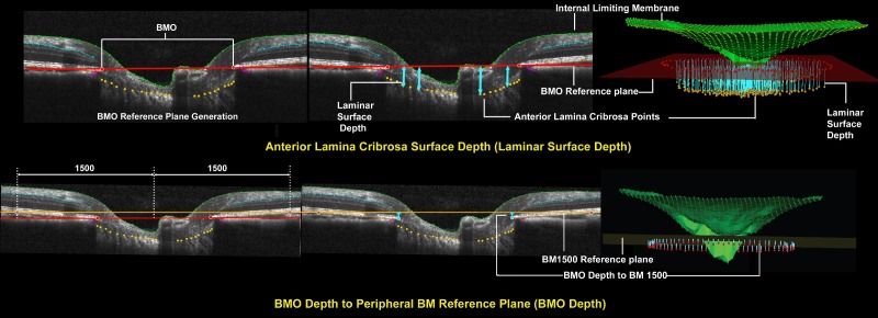
Figure 3.
Representative ONH connective tissue parameters within each B-scan and in 3D space (set 1). Top left: Bruch's membrane opening reference plane is defined using a best fit ellipse that fit through all 80 delineated BMO points (two in each radial). Top middle: Anterior lamina cribrosa surface depth (light blue arrows) is measured at each delineated anterior lamina cribrosa point as the perpendicular distance from the BMO reference plane (laminar surface depth) seen in cross-section as a red line. It is positive when anterior lamina cribrosa is above or anterior to the reference plane and negative when anterior lamina cribrosa is below or negative to the reference plane. Top right: Anterior lamina cribrosa depth in 3D space for a full 3D volume. Bottom left: Bruch's membrane reference plane definition. The BM reference plane is constructed in two steps. First, to identify constituent BM points, a circle within the BMO reference plane 1500 μm from the BMO centroid is identified and perpendiculars are dropped to the RPE/BM complex, in 7.5° intervals. Second, a best-fitting ellipse is fit through the identified RPE/BM complex points. Bruch's membrane opening depth is positive when BMO is above or anterior to the BM reference plane and negative when BMO is below or negative to the BM reference plane. Bottom middle: Bruch's membrane opening depth is measured at each delineated BMO point as the perpendicular distance from the BM reference plane (orange line) (bottom right).

