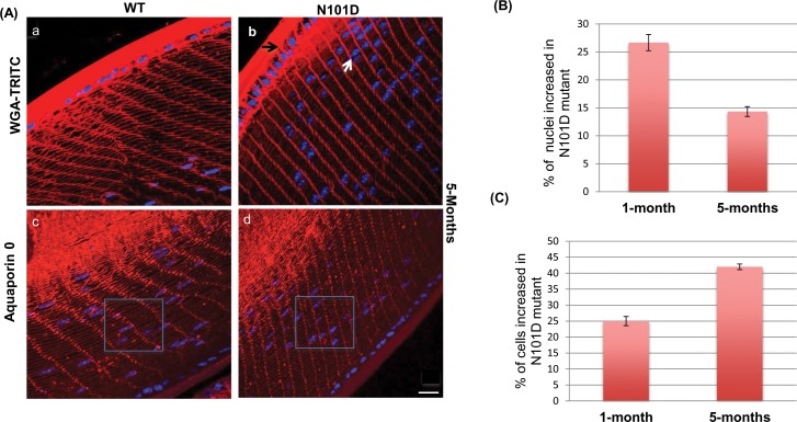
Figure 2.
Visualization of cellular assembly and organization in WT and N101D lenses by membrane labeling with WGA-TRITC and aquaporin-0 (A). Five-month-old equatorial sections were stained with WGA-TRITC (a, b), antiaquaporin 0 antibody (c, d), counterstained the nuclei with DAPI (blue). The black arrow in (b) shows increased thickness of epithelium in N101D compared to WT (a). The white arrow in N101D shows differences in nuclei in N101D compared to WT. Inset square in (c, d) demonstrate an increased cell number in N101D compared with WT lenses. Scale bar: 60 μm. (B) Quantification of increased number of nuclei in N101D relative to WT. Total number of nuclei in WT versus N101D was counted and quantified. Results are based on four lens sections from two mice for each age. (C) Quantification of increased number of cells in N101D relative to WT. Total number of cells inside the square were counted and quantified. The represented data are from four lens sections of two mice of each age.

