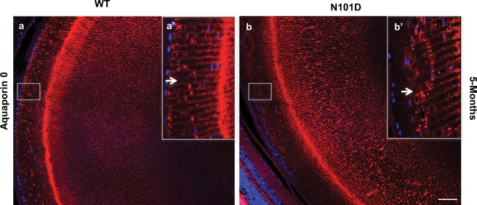
Figure 3.
Expression of aquaporin-0 is delayed in N101D mutant lenses. In WT lenses, the aquaporin-0 staining with antiaquaporin antibody was observed in transitional zone just bellow epithelium layer (a) and in (Inset a': enlarged area, indicated by an arrow in a'. In contrast, in the N101D mutant lenses, aquaporin-0 staining (b) and (Inset [b']: enlarged area) was observed farther away from the epithelial layer. Nuclei were counterstained with DAPI (blue). The results shown are from lens sections from two animals per age group. Scale bar: 80 μm.

