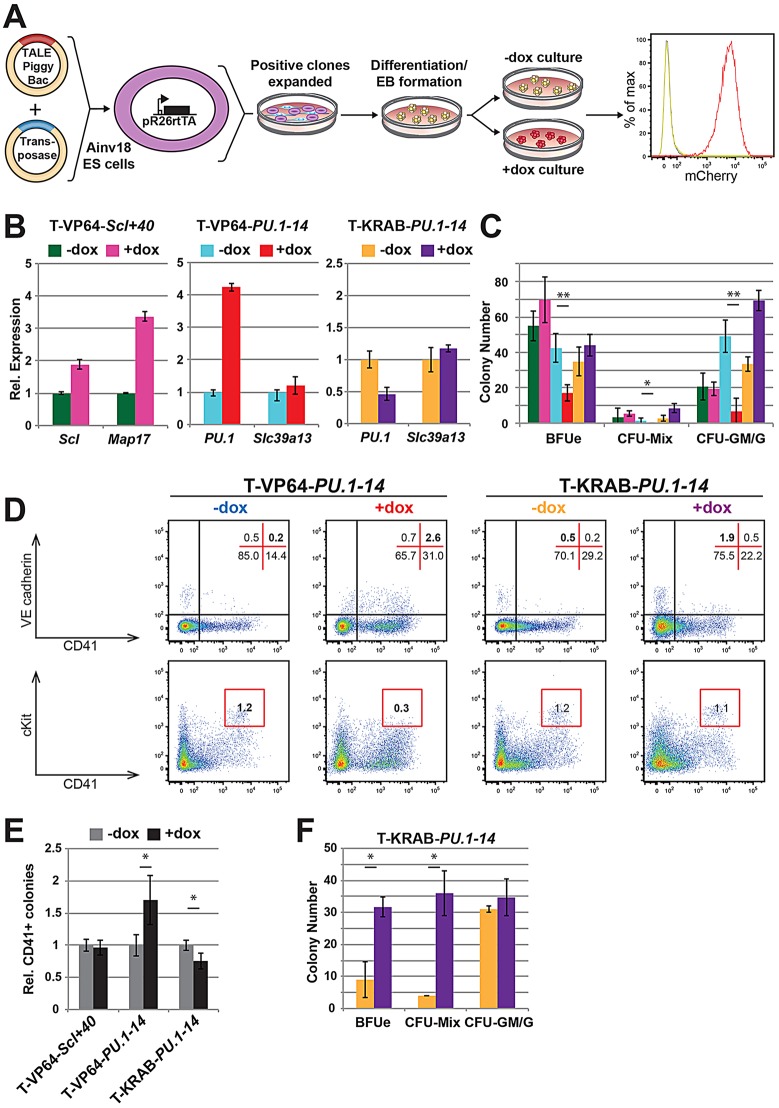
Fig. 2.
Transient TALE expression affects haematopoietic cell fate decisions. (A) Experimental approach using Ainv18 ESC differentiation to study TALE-mediated gene expression perturbations in haematopoiesis. Mouse Ainv18 ESCs constitutively expressing rtTA from the Rosa26 locus (pR26-rtTA) were co-transfected with the inducible TALE-PB construct and transposase. Targeted ESCs were differentiated into embryoid bodies (EBs), and TALE expression induced at day 4 by addition of dox. Changes in gene expression, colony potential and surface marker phenotype were analysed at day 6 in the +dox EBs as compared with −dox controls. (B) Gene expression changes in day 6 EBs after induction of T-VP64-Scl+40 (left), T-VP64-PU.1-14 (middle) and T-KRAB-PU.1-14 (right). Error bars indicate s.e.m. of three biological replicates. (C) Representative haematopoietic colony numbers from 1×10⁵ day 6 EB cells (colour scheme as in B). Colonies were grown in methylcellulose supplemented with SCF, IL-3, IL-6 and Epo. See supplementary material Fig. S2A for images of representative colony forming units (CFUs) scored. Error bars indicate s.d. of technical triplicates. *P<0.05, **P<0.01 (Student's t-test), from three biological replicates. (D) Flow cytometry plots of day 6 EB cells showing Flk1 versus CD41 (top) and VEcad versus CD41 (bottom). Representative staining patterns are shown for T-VP64-PU.1-14 (left) and T-KRAB-PU.1-14 (right). The distribution of cells within quadrants/gates is shown by percentage. (E) Relative number of day 4 Flk1+ EB-derived colonies containing CD41+ haematopoietic cells, grown on OP9 stromal cells for 84 h (dox added after 36 h). See supplementary material Fig. S2G for representative image of scored colony. Error bars indicate s.e.m. from biological triplicates. *P<0.01 (Student's t-test), from three biological replicates. (F) Average numbers of haematopoietic colonies from 1×10⁵ day 6 EB T-KRAB-PU.1-14 cells plated onto confluent OP9 stromal cells for 24 h before CFU assay initiated by addition of methylcellulose supplemented with SCF, IL-3, IL-6 and Epo. Colour scheme as in B. Error bars indicate s.d. of three biological replicates. *P<0.05 (Student's t-test), from three biological replicates.

