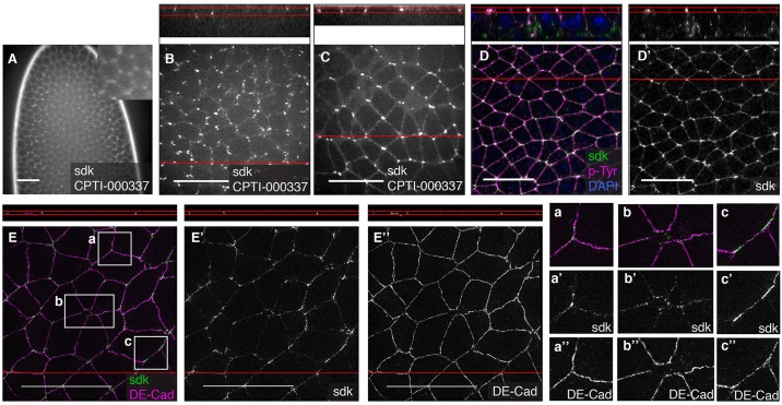
Fig. 5.
Localisation of Sidekick at tricellular vertices. (A,B) In live embryos at stage 5, CPTI-000337 inserted in Sidekick localises in spots that become progressively enriched apically, at cell vertices. (C) In live embryos at stage 8, Sidekick-YFP is localised apically and marks cell vertices between three or more cells. (D,D′) In fixed embryos of same stage, co-staining with p-Tyr confirms the apical junctional position of Sidekick-YFP. (E-E″) Superresolution imaging of fixed embryos at stage 8 stained for DE-Cadherin and YFP shows that DE-Cadherin and Sidekick tend to localise in complementary domains, with DE-Cadherin mainly at bicellular contacts (E,E″,a″,b″) and Sidekick (E,E′) at tricellular (a′) or multicellular contacts (b′). Occasionally, Sidekick forms plaques at bicellular contacts where Cadherin is less enriched (c′,c″). Top panels show side views from the reconstruction of the z planes at the position of the red line in the main images. Scale bars: 20 μm.

