
Fig. 2.
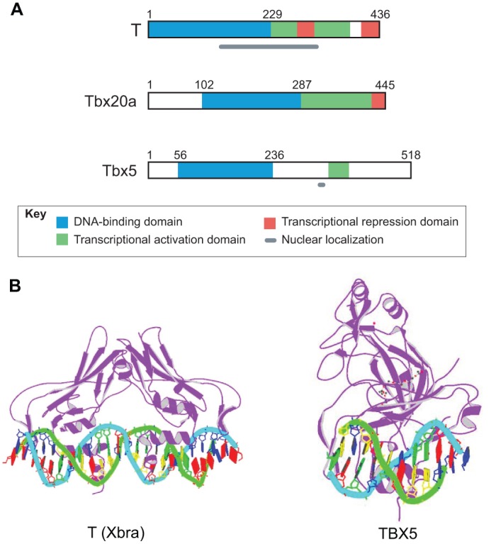
Domain structure of T-box proteins and crystal structure of T-domains bound to DNA. (A) The domain structure of three example T-box proteins [T (Kispert, 1995), Tbx20a (Stennard et al., 2003) and Tbx5 (Zaragoza et al., 2004)] illustrating the location of the DNA-binding domain in the N-terminal portion and either a transcriptional activation domain or activation and repression domains in the C-terminal region. The domain involved in nuclear localization has not yet been determined for Tbx20. (B) Crystal structure of T-domains from Xenopus laevis T (Xbra) bound to a palindromic T-box binding element derived from the in vitro selected consensus sequence (Müller and Herrmann, 1997) and from human TBX5 bound to a natural T-box binding site from the ANF promoter (Stirnimann et al., 2010). Images are from the RCSB Protein Data Bank (www.rcsb.org): ID numbers 1XBR (Xbra) and 2X6V (TBX5).