Figure 1.
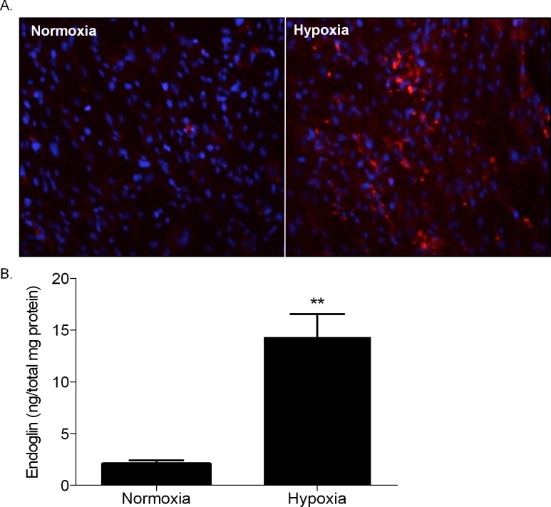
Endoglin induction by hypoxia. (A) Anti-CD105 immunocytochemistry in rat RMECs cultured in normoxia and hypoxia for 24 hours. Endoglin is shown in red, while DAPI-stained cell nuclei are shown in blue. (B) Endoglin (CD105) ELISA on human retinal microvascular endothelial cells exposed to normoxia and hypoxia for 24 hours. Endoglin levels are significantly upregulated in rat and human microvascular endothelial cells cultured in hypoxia versus normoxia. **P < 0.02, relative to normoxia).

