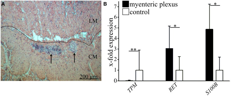
Figure 1.
Identification of enteric ganglia of human myenteric plexus and validation of laser microdissection technique. (A) Histological staining of large intestine with hematoxylin and eosin. Ganglia (marked by arrows) are visible between the outer longitudinal (LM) and the inner circular muscle (CM). Scale bar 200 μm. (B) Representative validation of laser microdissection technique by qRT-PCR in one donor. Black bars indicate the relative gene expression of tropomyosin (TPM1, smooth muscle marker, **P ≤ 0.005), Ret receptor (RET, neuronal marker, *P ≤ 0.05) and S100 calcium binding protein B (S100B, glial marker, *P ≤ 0.05) compared to full wall tissue controls (white bars). Data demonstrate that TPM is almost absent in microdissected myenteric plexus whereas Ret and S100 beta are both significantly enriched.

