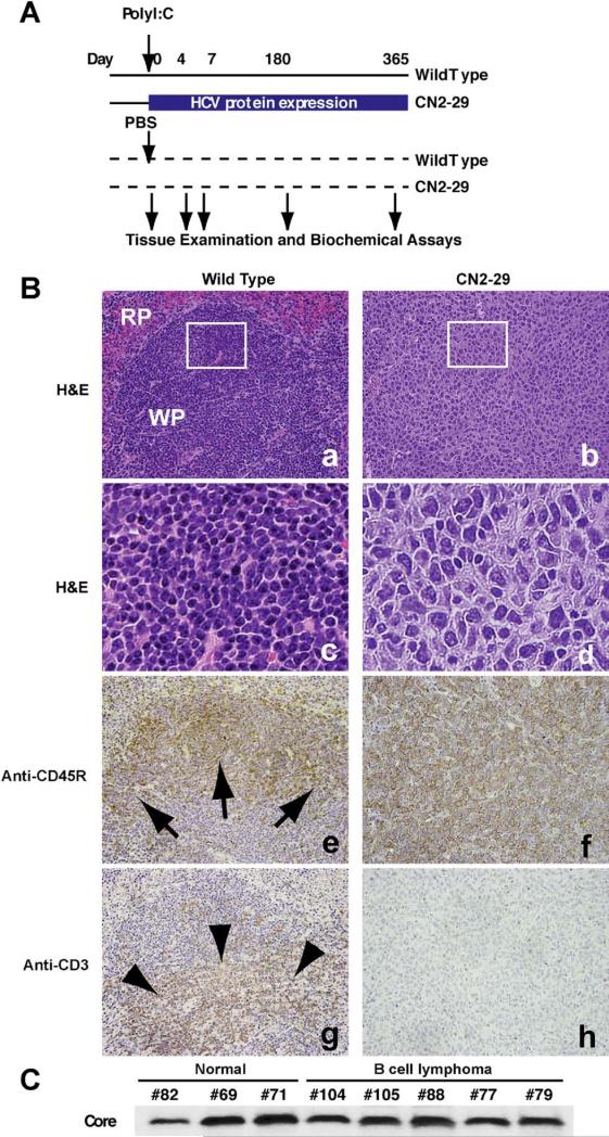
Figure 4.
Stable expression of HCV viral proteins induces lymphoproliferative diseases. (A) Switching of the expression of HCV proteins was conducted using the Mx promoter–driven cre recombinase with poly(I:C) induction. The Mx promoter is active in hepatocytes as well as in hematopoietic cells. We crossed CN2 mice with Mx1-Cre transgenic mice; Cre recombinase was expressed from the IFN-inducible Mx1 promoter. Injection of Mx1-Cre/CN2-29 mice with poly(I:C) induces IFN production and efficiently induces the expression of CN2 gene products in hematopoietic cells (mainly in Kupffer cells and lymphocytes), livers, and spleens but not in most other tissues. (B) The white pulp (WP) and red pulp (RP) comprise the components of the spleen in WT mice. The neoplastic cells replace the normal structures, such as the white pulp and red pulp. (c and d) The neoplastic cells are larger than lymphocytes (c), and the nuclei are irregular, round, oval, elon-gated, and polygonal (d). (e and g) The white pulp in WT mice consists of both a B-cell–rich area (arrows, e) and T-cell– rich area (arrowheads, g). (f and h) The neoplastic cells show staining for the B-cell marker CD45R, thereby supporting the diagnosis of B-cell lymphoma (f), while they do not show staining for the T-cell marker CD3 (h). Frames c and d are higher-magnification views of the white box areas in a and b, respectively. (C) Core protein expression was confirmed by immunoblotting.

