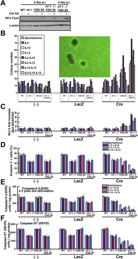
Figure 6.
Lymphocyte transformation by aberrant cytokines and inhibition of apoptotic signaling. (A) Expression of the HCV core protein (21 kilodaltons) in irf-1+/+ (WT) and irf-1−/− transgenic (CN2-29) and WT mice 2 or 4 days postinfection (p.i.) with AxCANCre (multiplicity of infection, 1.0). β-Actin was used as a loading control. (B) Colony formation assay for splenocytes from irf-1+/+ (WT) and irf-1−/− WT or transgenic (CN2-29) mice in the absence or presence of the indicated cytokine and infected with mock, LacZ, and Cre adenoviruses. The inset shows an image of the colonies generated from the irf-1−/− CN2 splenocytes (original magnification 10×). (C) Quantification, by quantitative reverse-transcription PCR of Bcl-2 mRNA relative to control glyceraldehyde-3-phosphate dehydrogenase mRNA in the splenocytes of irf-1+/+ (WT) and irf-1−/− or transgenic (CN2-29) mice treated with the indicated cytokines and infected with mock, LacZ, and cre adenoviruses. (D) Apoptosis measured by Annexin V fluorescence-activated cell sorting analysis of splenocytes from irf-1+/+ (WT) and irf-1−/− or transgenic (CN2-29) mice treated with the indicated cytokines and infected with the mock, LacZ, and cre adenoviruses. (E and F) The caspase-9 and caspase-3/7 enzymatic activities in splenocytes from irf-1+/+ (WT) and irf-1−/− or transgenic (CN2-29) mice treated with the indicated cytokines were measured using a substrate cleavage assay after infection with the mock, LacZ, and Cre adenoviruses. Caspase-9 activity was measured 4 hours after injection of the anti-Fas monoclonal antibody (Jo2). LEHD, substrate for caspase-9; DEVD, substrate for caspase-3/7. Vertical bars are SD and were determined using the Student t test.

