
Figure 1.
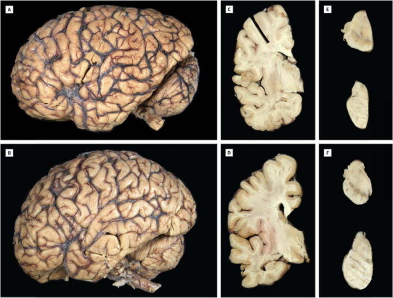
Macroscopic features of the C9ORF72-associated pseudodementia patients. (A, B) Fixed left hemibrain of the pseudodementia patients both show no cortical atrophy. Coronal sections show a thick cortical ribbon, corpus callosum and a normal appearing thalamus and hippocampus with no ventricular enlargement in Patient 1 (C) and minimal enlargement in Patient 2 (D). (E, F) There is visible pigment in the substantia nigra (midbrain; upper) and locus ceruleus (pons; lower) of both individuals.