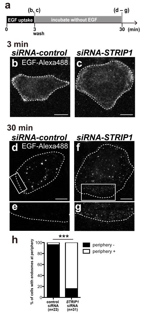
Figure 6.
Localization defects of EGF-Alexa488 in STRIP1 knockdown HeLa cells
(a) Summary of EGF-Alexa488 uptake assay. (b–g) Localization of EGF-Alexa488 in the control (b,d,e) and STRIP1 (c,f,g) siRNA-treated HeLa cells. HeLa cells were transfected with control or STRIP1 siRNA for 3 days, and endocytosis uptake was assayed with EGF-Alexa488. After a 3 min uptake of EGF-Alexa488, cells were fixed (b,c) or washed with DMEM and cultured for 27 min and fixed (d–g). The rectangle regions of (d) and (f) are magnified at (e) and (g), respectively. Scale bar: 10 μm. (h) All cells were classified into having or not having EGF-Alexa488 at cell periphery by a blind test. ***p<0.0001, chi-square test.

