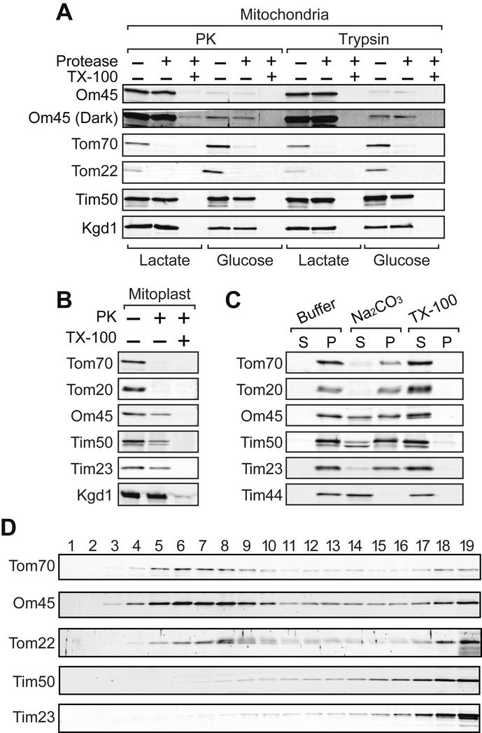
Figure 1. Om45 is an OM protein exposed to the IMS.

A Mitochondria isolated from the cells cultured in YPD or lactate medium were treated with 125 μg/ml of PK or trypsin in the presence or absence of 0.2% Triton X-100 for 20 min on ice. Proteins were analyzed by SDS–PAGE and immunoblotting with the indicated antibodies. In Om45 (Dark), contrast of the image was enhanced.
B Mitoplasts were PK-treated, and proteins were analyzed as in (A).
C Mitochondria were incubated in SEM buffer (Buffer), with 0.1 M Na2CO3 or with 1% Triton X-100 (TX-100), on ice for 20 min and then subjected to ultracentrifugation. The resulting supernatants and pellets were analyzed as in (A).
D OM and IM vesicles generated from mitochondria were subjected to sucrose density-gradient centrifugation. Each fraction was analyzed as in (A).
