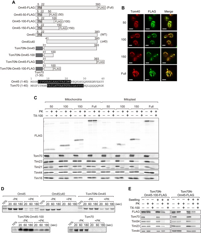
Figure 2. The N-terminal 50 residues direct Om45 to the IMS.
A Top: A schematic diagram of Om45 variants (with the FLAG tag). Bottom: The predicted TM segments in residues 1–40 of Om45 and Tom70 are indicated by white letters on black background.
B Localization of the Om45 variants was assessed by immunofluorescence with the anti-FLAG antibody (FLAG). The mitochondria were visualized with anti-Tom40 antibodies (Tom40). Scale bar, 3 μm.
C Mitochondria or mitoplasts with the indicated Om45 variants were incubated with 125 μg/ml PK with or without 0.2% Triton X-100 (TX-100) for 20 min on ice. Proteins were analyzed by SDS–PAGE and immunoblotting with the indicated antibodies.
D The indicated 35S-labeled proteins were imported into mitochondria in vitro for the indicated times and PK-treated. Imported proteins were analyzed by SDS–PAGE and radioimaging.
E Mitochondria (−Swelling) or mitoplasts (+Swelling) with the indicated Om45 variants were analyzed as in (C).

