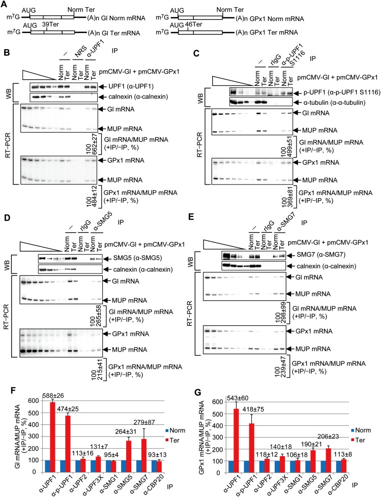
Figure 2.
p-UPF1, SMG5, and SMG7 preferentially bind to PTC-containing mRNAs compared with their PTC-free counterparts. (A) Diagrams of spliced Gl and GPx1 PTC-free (Norm) and Gl and GPx1 PTC-containing (39 Ter and 46 Ter, respectively) mRNAs. Boxes represent coding regions, vertical lines within boxes show spliced junctions, and horizontal lines denote UTRs. (B) HEK293T cells (8 × 10⁷ per 150-mm dish) were transiently transfected with 2 μg of phCMV-MUP and either 4 μg of pmCMV-Gl Norm + 4 μg of pmCMV-GPx1 Ter or 4 μg of pmCMV-Gl Ter + 4 μg of pmCMV-GPx1 Norm. Immunoprecipitations of lysates were performed using anti-UPF1 (α-UPF1). (Top) Western blotting (WB) before (−) or after immunoprecipitation (IP) using anti-UPF1 or, as a control for nonspecific immunoprecipitation, normal rabbit serum (NRS), where lanes below the wedge analyze serial threefold dilutions of lysate. (Bottom) RT–PCR, where the level of Gl mRNA or GPx1 mRNA before and after immunoprecipitation was normalized to the level of MUP mRNA, the normalized level after immunoprecipitation was calculated as a ratio of the normalized level before immunoprecipitation, and the ratio for Gl Norm mRNA or GPx1 Norm mRNA is defined as 100%. Lanes below the wedge analyze serial twofold dilutions of lysate RNA. (C) As in B, only immunoprecipitation was performed using anti-p-UPF1(S1116) or, as a control, rIgG. (D) As in B, only immunoprecipitation was performed using anti-SMG5. (E) As in B, except immunoprecipitation was performed using anti-SMG7. (F) RT-qPCR of Gl mRNA from samples analyzed in B–E and Supplemental Figure S2. (G) As in F for GPx1 mRNA. All quantitations derive from three to four independently performed experiments and represent the mean plus standard deviations.

