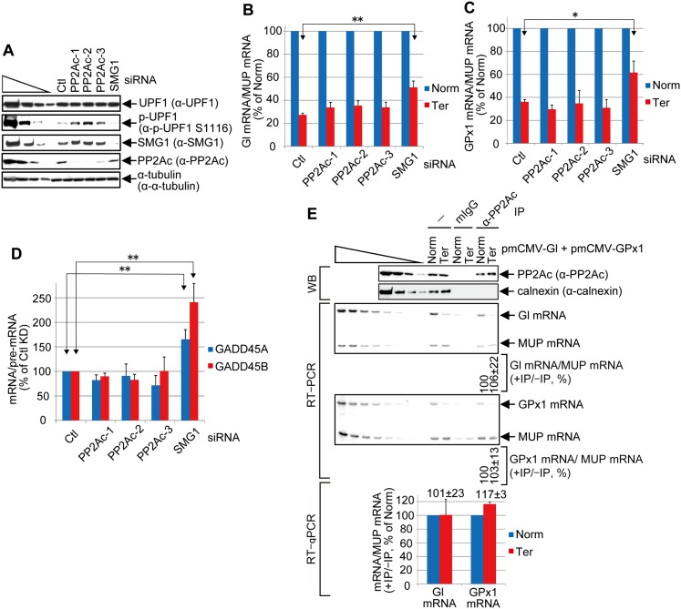
Figure 6.
Evidence that PP2Ac may dephosphorylate p-UPF1 after mRNA decay initiates. (A) Western blotting of lysates of HEK293T cells (5 × 10⁶ per each six-well plate) transiently transfected with 60 pmol of the specified siRNA and, 1 d later, 0.4 μg of pmCMV-Gl Norm or Ter, 0.4 μg of pmCMV-GPx1 Norm or Ter, and 0.2 μg of phCMV-MUP. (B) Using samples from A, RT-qPCR of Gl Norm or Ter mRNA as described in Figure 2F for Gl mRNA before immunoprecipitation. (C) As in B but analyzed as in Figure 2G for GPx1 mRNA before immunoprecipitation. (D) Using samples from A, RT-qPCR of endogenous NMD targets GADD45A and GADD45B mRNAs. mRNA levels were normalized to the level of the corresponding pre-mRNA using PCR primers described in Supplemental Table S2. Quantitations in B–D derive from three independently performed experiments and represent the mean plus standard deviations as statistically analyzed using the two-tailed t-test. (*) P < 0.05; (**) P < 0.01. (E) As in Figure 2, B, F, and G, but using anti-PP2Ac or mouse IgG (mIgG) in the immunoprecipitations. Two to three independently performed experiments represent the mean plus standard deviations.

