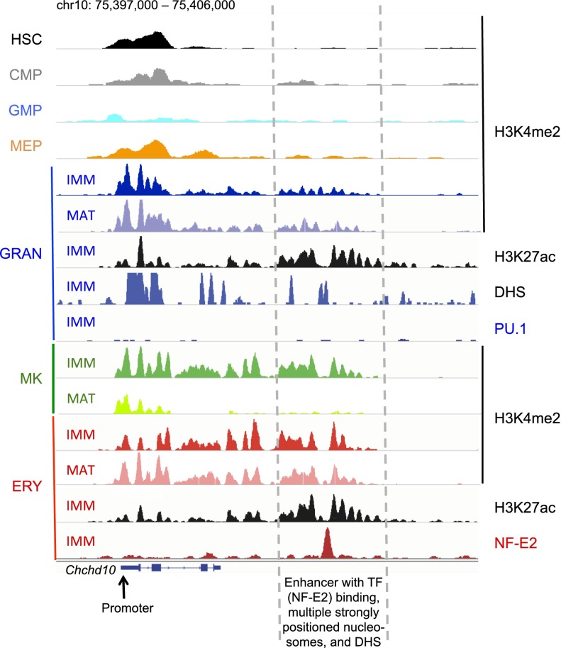
Figure 6.
Chronology and infidelity of enhancer delineation at a representative locus illustrate the principal conclusions of this study. IGV traces at a representative erythroid-expressed locus, Chcd10, show strong NF-E2 binding at a distant erythroid enhancer (dashed lines) that does not bind PU.1 in granulocytes. H3K4me2-marked nucleosomes flank this enhancer in erythroid cells and MKs, and weakly in granulocytes, with significant resolution of the mark in terminally mature MKs. This region carries strong H3K27ac marks and DHS in granulocytes, indicating enhancer delineation. The promoter (arrow) is strongly marked in most blood cells, but enhancer marks are extremely weak to absent in HSC or progenitor cells.

