Figure 4.
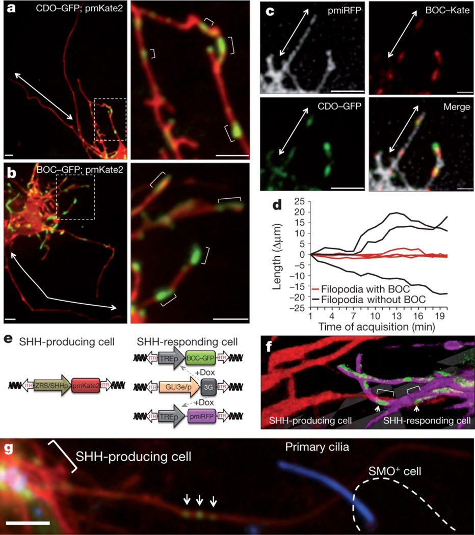
Filopodia on Shh responding cells display an exquisite distribution and co-localization of Shh co-receptors that interact with Shh producing filopodia. (a1) Live imaging of Cdo-GFP and (b1) Boc-GFP expression in defined microdomains along the filopodial membrane, within subsets of filopodia but not others (arrows). (a2–b2) Higher magnification of Fig. a1 and b1 showing multiple positive microdomains of co-receptor localization (brackets) interspersed along the filopodia membrane. (c1–4) Cdo-GFP and Boc-Kate2 are colocalized along microdomains of filopodia (arrow) labeled with membrane-associated near-infrared fluorescent protein (pmiRFP). (d) Boc-harboring filopodia (red line) are statistically more stabilized than filopodia without Boc (black line), p < 0.001, n=160 timepoints. (e) Expression system to specifically label Shh producing cells and Shh responding cells in the same limb bud. (f) Representative 3D image of a filopodia from a Shh producing cell (pmKate2 - red) that interacts with domains of Boc-GFP (green) along the filopodia membrane (pmiRFP, fuscia) of Shh responding cell. Arrows show interaction along Boc microdomains (brackets). (g) Shh producing cell, indicated by pmKate2 and marked by bracket, with a long filopodium containing ShhN-EGFP particles (arrows) that contacts a Smoothened positive cell, Smo+, (outlined by a dashed line). Smoothened-BFP localization to the cilium is a marker of Shh pathway activation. Scale = 3µm.

