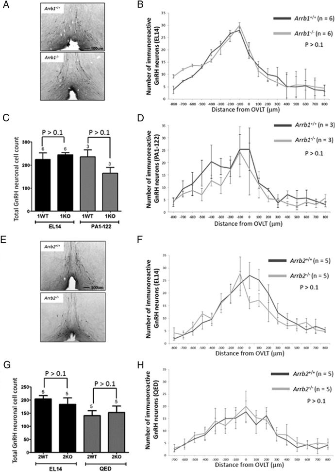
Figure 1.
Loss of either Arrb1−/− or Arrb2−/− has no effect on the GnRH-immunoreactive neuronal population size and distribution in the hypothalamus. A and E, Photomicrographs of coronal brain sections at the level of the OVLT from 8-week-old Arrb1−/− and Arrb2−/− mice and their WT littermate. In photomicrographs, GnRH neurons were revealed in the brain slices by immunohistochemistry using the EL14 antibody, and the OVLT was used as a neuroanatomical reference point for rostral to caudal alignment of coronal sections. B, D, F, and H, Histograms displaying the rostral-caudal distribution of GnRH-immunoreactive neurons from WT and KO mice aligned at the OVLT. The plot represents average GnRH-immunoreactive neurons per 50-μm coronal section. GnRH-immunoreactive neurons were identified with the EL14 antibody (B, Arrb1−/− mice; F, Arrb2−/− mice), PA1–122 antibody (D, Arrb1−/− mice), and QED antibody (H, Arrb2−/− mice). C and G, Total number of GnRH-immunoreactive neurons identified with the EL14 and PA1–122 antibody (C, Arrb1−/− mice) and EL14 and QED antibody (G, Arrb2−/− mice). Error bars represent SEM.

