Figure 4.

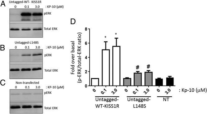
L148S triggers ERK1/2 phosphorylation. A–D, Representative autoradiographs (A–C) and densitometric analysis (D) showing the expression of total and phosphorylated ERK1/2 in WT untagged KISS1R- and untagged L148S-overexpressing HEK 293 cells and nontransfected (NT) HEK 293 cells after a 10-minute Kp-10 treatment (0μM, 0.1μM, and 3.0μM). Western blot analyses were done using monoclonal anti-ERK1/2 and anti–phospho-ERK1/2 antibodies. The data represent the mean of 3 independent experiments. *, P < .05, pERK1/2/total ERK1/2 ratio compared with 0μM in WT group; #, P < .05, pERK1/2/total ERK1/2 ratio compared with 0μM in L148S group. Error bars represent SEM.
