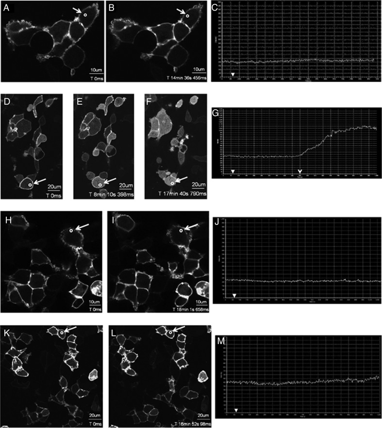
Figure 8.
L148S is strongly uncoupled from Gαq/11 signaling. A–M, representative images selected from a time series of laser scanning confocal microscopic images showing the plasma membrane and cytosolic localization of GFP-PH-PLC-δ1 in nontransfected HEK 293 cells (A–C) and in HEK 293 cells expressing WT KISS1R or L148S (D–G, FLAG-WT-KISS1R-expressing cells; H–J, FLAG-L148S-expressing cells; K-M: myc-L148S-expressing cells) in the absence of agonist (0 seconds) and in response to 3μM Kp-10 treatment (added gently and in a drop-wise manner to cells cultured on a confocal dish) approximately 1 minute later (see solid white arrowheads on x-axis of graphs C, G, J, and M). Cytosolic regions of interest in cells (shown by small white circle in panels A, B, D–F, H, I, K, and L) were analyzed quantitatively by determining the changes in GFP fluorescence over time. These changes are presented graphically (C, G, J, and M). These studies were conducted 5 independent times, and representative images from 1 independent experiment are shown. A–C, Approximately 14 minutes after Kp-10 treatment, translocation of GFP-PH-PLC-δ1 from the plasma membrane to the cytosol was visually undetectable. This confirms that translocation is dependent on KP/KISS1R signaling. D–G, At approximately 440 seconds (7.5 minutes) after adding Kp-10 (open arrowhead) GFP-PH-PLC-δ1 began translocating from the plasma membrane to the cytosol. Thus, the KISS1R-coupled Gαq/11 pathway is activated. H–J, Translocation of GFP-PH-PLC-δ1 from the plasma membrane to the cytosol was visually undetectable after 15 minutes of Kp-10 treatment. K–M, Translocation of GFP-PH-PLC-δ1 from the plasma membrane to the cytosol was visually undetectable after 16 minutes of Kp-10 treatment.

